
02 · During trial
Run imaging with full auditability
- Image receipt, completeness and quality control
- Visual reads by trained experts, for efficacy and safety
- Quality management (ISO 9001:2015)
Imaging · Machine learning · Clinical trials
QSA applies the latest imaging and machine learning technologies to show more precisely where treatments are working, across every stage of a clinical trial.
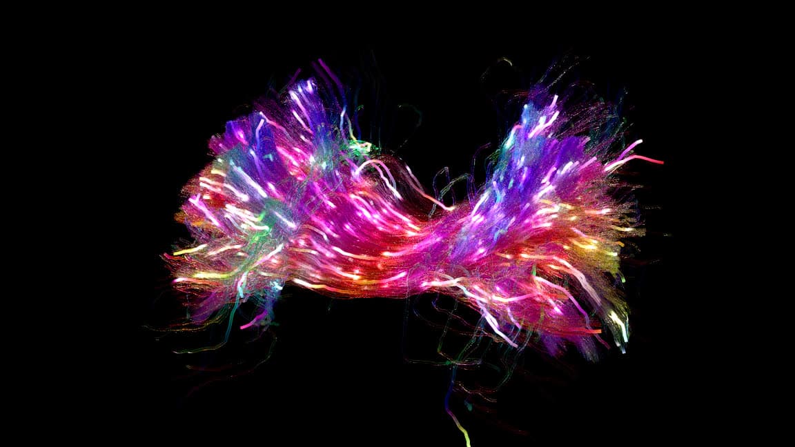
Corpus callosum tractography
Services along the trial life cycle

01 · Pre-trial
02 · During trial

03 · Post-trial
 (opens ARIAEducation.eu in a new tab)
04 · Imaging education and consulting
Education and specialised studies
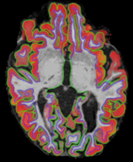
Technology and biomarkers
Machine learning that flags image issues such as blurring, ghosting and banding.
Processing pipelines with integrated quality control and interactive scan viewing.
Expert review with multi-visit comparison, lesion annotation and safety reads.
Objective measurement of brain volume percentage change.
Deep learning for rapid brain-atrophy outcomes in phase 2 and 3 trials.
Data-driven disease subtypes, with each patient staged within them.
Areas of work
MS subtypes and stages from MRI and blood biomarkers, found with SuStaIn.
Willard et al., Brain 2025 (opens in new tab)MRI analysis for a trial of CAR-T cell therapy in progressive MS.
ARIAEducation.eu, training in recognising ARIA on MRI.
ARIAEducation.eu (opens in new tab)Every scan checked for artefacts on arrival.
Ravì et al., Medical Image Analysis 2024 (opens in new tab)DeepBrainPrint finds scans of the same person across visits and scanners.
Puglisi et al., MIDL 2024 (opens in new tab)Subtypes and stages of diabetic retinopathy, found with SuStaIn.
Evidence
A multi-year MS subtyping study through image analysis.
A three-year imaging analysis for an MS trial of a CAR-T cell therapy.
Tell us about your study and the imaging questions behind it.